- Case report
- Open access
- Published:
A case report of Klebsiella aerogenes-caused lumbar spine infection identified by metagenome next-generation sequencing
BMC Infectious Diseases volume 22, Article number: 616 (2022)
Abstract
Background
The early clinical diagnosis of spinal infections in elderly patients with recessive or atypical symptoms is difficult. Klebsiella aerogenes is a common opportunistic bacterium that can infect the respiratory tract, urinary tract, and even the central nervous system. However, whether it can infect the lumbar spine has not been previously described.
Case presentation
In this paper, we report the case of a 69-year-old female patient with osteoporosis who was initially diagnosed with hemolytic anemia. Later, she was diagnosed with K. aerogenes infection of the lumbar spine based on imaging combined with blood culture and metagenome next-generation sequencing (mNGS) detection. After precise medication, the lumbar degeneration was improved.
Conclusions
Bacterial infection should therefore be considered in cases of lumbar degenerative disease in middle-aged and elderly patients.
Background
Klebsiella aerogenes, formerly known as Enterobacter aerogenes, belongs to the family Enterobacteria and is a facultative Gram-negative anaerobe [1]. It is widely distributed in the environment and is found in the human gastrointestinal tract, also being a common opportunistic pathogen in hospitals. When the host immune system is compromised or the intestinal mucosa is damaged, it may cause infection of the respiratory, circulatory, or urogenital system [2]. In recent years, despite increasing reports on the pathogenicity and drug resistance of Escherichia coli and Klebsiella pneumoniae, there have only been a few reports on K. aerogenes [3, 4]. Previous clinical reports on this bacterium were mainly of respiratory tract, gastrointestinal tract, urinary tract, and blood infections. Compared with other Enterobacteriaceae species, K. aerogenes is more likely to cause septic shock or even death in patients[5, 6]. To date, lumbar infections caused by K. aerogenes have not been reported. Herein, we report the first case of a patient with a lumbar K. aerogenes infection.
Case presentation
A 69-year-old woman was hospitalized on July 30, 2020, with recurrent fever without any cause for two weeks and continuous lower back pain for three days. Three months prior, she presented with chest discomfort and fatigue without an obvious cause, occasional chest pain after activity, and frequent urination at night. She did not have paroxysmal nocturnal dyspnea, fever, cough, expectoration, urinary pain, nausea, vomiting, abdominal pain, abdominal distension, or diarrhea. Physical examination revealed high inflammatory indices, including a white blood cell (WBC) count of 21.7 × 10⁹ cells/L, a C-reactive protein (CRP) level of 12.46 mg/L, and procalcitonin (PCT) level of 0.19 ng/mL. She was treated for infection and anemia, as laboratory tests revealed an erythrocyte count of 1.00 × 10¹²/L, a hematocrit of 14.0%, a hemoglobin concentration of 43 g/L, a mean corpuscular volume (MCV) of 140.0 fL, a mean corpuscular hemoglobin (MCH) level at 43.0 Pg, and a MCH concentration of 307 g/L (Table 1). The symptoms did not improve, and the patient remained at our hospital for treatment.
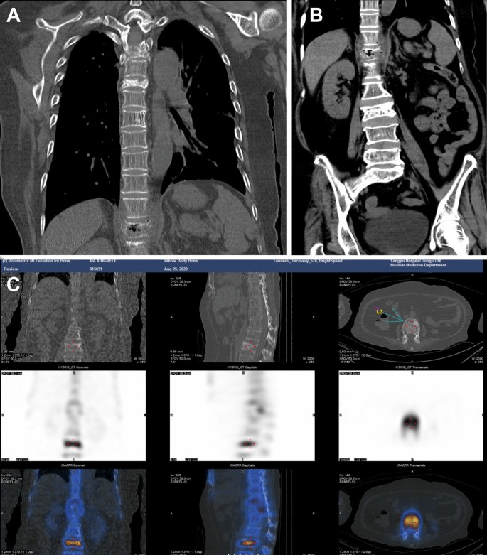
The chief complaint at admission: In 2006, the patient underwent radical mastectomy and chemotherapy after surgery, with a 7-year history of hypertension and continuous lower back pain for three days. Through physical examination, it was found that the patient had back tenderness and paravertebral percussion pain, leading to the diagnosis of thoracic degenerative changes, particularly compression changes at the fifth and the twelfth thoracic as well as the third lumbar vertebra. Body temperature fluctuated between 38.6 and 38.8 °C, and the patient was treated with ceftriaxone and ceftazidime. On August 18, she was hospitalized again due to recurrent fever for 2 weeks and severe diarrhea for 1 day. Chest computed tomography (CT) revealed the absence of soft tissue shadows in the right breast and right armpit, increased texture in both lungs, a cord-like shadow in both lungs, a small amount of left pleural effusion, and bilateral pleural thickening (Fig. 1A).
Abdominal CT showed a slightly enlarged spleen, local pneumatosis expansion in the proximal colon ascendens, and no other visible abnormalities (Fig. 1B). Blood cultures were positive for K. aerogenes within 15 h (Table 2), and the result of urine cultures was negative, while stool cultures were positive for Candida albicans and negative for Enterobacter sp. and Vibrio casei. Based on drug sensitivity testing and the patient’s condition, meropenem and diflucan (fluconazole resistance) were administered against infection. Detailed information on the drug sensitivity testing was provided in the Additional file 1: Online Technical Appendix. After 4 days, body temperature was normal, and the patient was treated with sulperazon.
On August 25, the patient, who had a history of breast cancer, experienced chest discomfort. A bone scan with the whole-body bone scintigraphy was performed, confirming the absence of bone metastases, while also revealing cuneiform changes at the third, fifth, twelfth thoracic vertebrae, and the third lumbar vertebra, indicative of degenerative changes in the thoracolumbar spine (Fig. 1C). During follow-up from anemia treatment, the patient had a normal hemoglobin level and was administered oral prednisone at a dose of 10 mg bid. The patient’s D-dimer levels remained high, and an improved pulmonary artery CT angiography (CTA) suggested that the small branch was embolized in the lower lobe of both lungs. The patient was treated with low-weight molecular heparin and switched to oral anticoagulants (rivaroxaban) when under stable condition.
On August 27, the patient returned to a normal body temperature, and antibiotics were discontinued. On August 31, the patient had increased PCT level, so she was once again treated with sulperazon against infection until she was discharged on September 12. There was no significant change in the blood routine and PCT indices during this period. She experienced a fever again without an obvious cause one day after discharge. Upon admission, the highest body temperature was 39.5 °C, accompanied by aversion to cold, chills, and fatigue. Physical examination revealed a CRP level of 88.89 mg/L, a WBC count of 14.4 × 10⁹ cells/L, a body temperature of 40.4 °C, and shock. K. aerogenes infection was detected through blood cultures within 12 h, and the patient was subsequently treated with meropenem against infection.
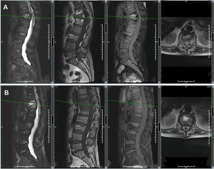
A CT scan of the thoracoabdominal region showed significant changes in the twelfth thoracic vertebra. Abdominal CT revealed vertebral compression change of the third lumbar vertebrae. MRI of the lumbar spine showed bone destruction from the eleventh thoracic vertebrae to the first lumbar vertebrae, protrusion of the intervertebral disc at the fifth lumbar vertebra to the first sacral vertebra, and degenerative changes in the lumbar spine (Fig. 2A ). Therefore, we considered that the patient had a vertebral infection. The patient experienced recurrent vague back pain, which she tolerated over half a year. Considering bone destruction, the expert consultation from another hospital led to the administration of cefepime combined with levofloxacin for the treatment of infection. One day later, the patient once again had a fever, and the axillary temperature was 38.5 °C, with a CRP level of 47.9 mg/L and a WBC count of 14.6 × 10⁹ cells/L. The antibiotic was changed to meropenem combined with moxifloxacin, and the following two times of blood cultures were negative.
On October 10, MRI of the lumbar spine revealed bone destruction from the eleventh thoracic vertebra to the first lumbar vertebrae. The thoracic MRI indicated mild deviation with signal changes of the eleventh thoracic vertebrae and the first lumbar vertebrae as well as vertebral compression with signal abnormalities of the twelfth thoracic vertebrae (Fig. 2B). From October 16 to 29, blood cultures were negative twice in a row, but K. aerogenes was detected via metagenomic next-generation sequencing (mNGS) on the 21st (Table 3). The specific methods of mNGS were described in the Additional file 1: Online Technical Appendix. Cefepime and moxifloxacin were administered continuously. The patient’s body temperature and inflammatory indexes tended to be normal during the treatment. After discharge, the patient continued oral administration of levofloxacin as well as doxycycline for infection treatment and was instructed to rest in bed to prevent falls, avoid stress on the waist, and appropriately move the lower limbs to avoid thrombosis. During the follow-up period, routine blood tests, inflammatory indexes, and body temperature were normal.
Discussion and conclusions
The present work describes case of a lumbar spine infection caused by K. aerogenes. The treatment process was complex, mainly due to the older age of the patient, the insignificant symptoms of lumbar infection, and no previous experience with spinal K. aerogenes infections. After repeated CT reexamination and communication with the patient about her physical symptoms, the therapeutic results were satisfactory. Based on our observations, we suggest that the possibility of spinal bacterial infection should be fully considered, and rare spinal pathogens, such as K. aerogenes, should be included in the diagnostic scope when treating elderly patients who develop lumbar degenerative diseases.
K. aerogenes, which has a thick capsule, belongs to the genus Klebsiella of Enterobacteriaceae. It is a Gram-negative facultative anaerobe that is widely distributed in the natural environment and animal intestines [7]. Like other Enterobacteriaceae pathogens, Klebsiella virulence and drug resistance are complex and influenced by multiple factors. The produced virulence factors and the drug resistance genes can vary depending on the site of infection. Adhesins produced by Klebsiella facilitate its entry into host cells, with capsular polysaccharides and lipopolysaccharides on the cell surface helping the bacteria to escape from the phagocytosis, while toxins or other extracellular components cause mucosal damage and spread through the circulation. In recent years, due to the excessive use of antibiotics, an increasing number of Klebsiella species have developed multidrug resistance [8]. As reported, carbapenem-resistant K. aerogenes isolates were previously found in clinical studies in China [9, 10]. In our present study, drug sensitivity test results indicated that K. aerogenes isolates were non-resistant bacteria to carbapenems, and were especially sensitive in meropenem, which was given as anti-infection treatment. In addition, the isolated K. aerogenes was intermediate in imipenem. Nevertheless, they were found to be resistant to penicillins, such as ampicillin and amoxicillin. Generally, Klebsiella-related infections develop rapidly, causing multiple organ failure or even death, and drug-resistant strains easily arise. Thus, effective antibiotics should be selected as early as possible, and the dose as well as course of treatment should be determined to minimize the occurrence of side effects.
Due to the lack of specific symptoms and signs, the early diagnosis of spinal infections is relatively difficult. Clinicians have to differentiate it based on characteristics of the disease, clinical symptoms, and signs, combined with X-ray, CT, MRI, and other imaging findings. Spinal infection is mostly subacute or chronic, and generally occurs in patients with weakened immune function or spinal surgery. In recent years, spinal infections have been reported to be caused by dental problems in adult patients with normal immunity [11]. Pain at the lesion site is the initial symptom, which may be accompanied by an elevated body temperature. Pathogens in patients with spinal infectious diseases are usually identified by blood culture. The common pathogens are Staphylococcus aureus, Streptococcus sp., Brucella sp., and Mycobacterium tuberculosis [12,13,14]. At present, there are no published case reports of K. aerogenes spinal infection. The patient in this study, an elderly female, was diagnosed with K. aerogenes infection of the lumbar spine based on imaging combined with blood culture and mNGS detection. After diagnosis and precise medication, lumbar degeneration was improved, and the patient was satisfied with the treatment. The present case highlights the possibility of spinal bacterial infection when lumbar degeneration is observed in middle-aged and elderly patients.
Availability of data and materials
The datasets used and/or analyzed during the current study are available from the corresponding author on reasonable request.
Abbreviations
- MNGS:
-
Metagenome next-generation sequencing
- WBC:
-
White blood cell
- CRP:
-
C-reactive protein
- PCT:
-
Procalcitonin
- CT:
-
computed tomography
- CTA:
-
Computed tomography angiography
- MCV:
-
mean corpuscular volume
- MCH:
-
Mean corpuscular hemoglobin
References
Wesevich A, Sutton G, Ruffin F, Park LP, Fouts DE, Fowler VG Jr, Thaden JT. Newly named Klebsiella aerogenes (formerly Enterobacter aerogenes) is associated with poor clinical outcomes relative to other Enterobacter Species in patients with bloodstream infection. J Clin Microbiol. 2020;58(9):e00582-20.
Li E, Wei X, Ma Y, Yin Z, Li H, Lin W, Wang X, Li C, Shen Z, Zhao R, et al. Isolation and characterization of a bacteriophage phiEap-2 infecting multidrug resistant Enterobacter aerogenes. Sci Rep. 2016;6:28338.
Pulcrano G, Pignanelli S, Vollaro A, Esposito M, Iula VD, Roscetto E, Soriano AA, Catania MR. Isolation of Enterobacter aerogenes carrying blaTEM-1 and blaKPC-3 genes recovered from a hospital Intensive Care Unit. APMIS. 2016;124(6):516–21.
Pereira RS, Dias VC, Ferreira-Machado AB, Resende JA, Bastos AN, Andrade Bastos LQ, Andrade Bastos VQ, Bastos RV, Da Silva VL, Diniz CG. Physiological and molecular characteristics of carbapenem resistance in Klebsiella pneumoniae and Enterobacter aerogenes. J Infect Dev Ctries. 2016;10(6):592–9.
Davin-Regli A, Pages JM. Enterobacter aerogenes and Enterobacter cloacae; versatile bacterial pathogens confronting antibiotic treatment. Front Microbiol. 2015;6:392.
Lin TY, Chi HW, Wang NC. Pathological fracture of the right distal radius caused by Enterobacter aerogenes osteomyelitis in an adult. Am J Med Sci. 2010;339(5):493–4.
Davin-Regli A, Lavigne JP, Pages JM. Enterobacter spp.: update on taxonomy, clinical aspects, and emerging antimicrobial resistance. Clin Microbiol Rev. 2019; 32(4).
Wolff N, Hendling M, Schroeder F, Schonthaler S, Geiss AF, Bedenic B, Barisic I. Full pathogen characterisation: species identification including the detection of virulence factors and antibiotic resistance genes via multiplex DNA-assays. Sci Rep. 2021;11(1):6001.
Shen Y, Xiao WQ, Gong JM, Pan J, Xu QX. Detection of New Delhi Metallo-Beta-Lactamase (Encoded by blaNDM-1) in Enterobacter aerogenes in China. J Clin Lab Anal. 2017; 31(2).
Qin X, Yang Y, Hu F, Zhu D. Hospital clonal dissemination of Enterobacter aerogenes producing carbapenemase KPC-2 in a Chinese teaching hospital. J Med Microbiol. 2014;63(Pt 2):222–8.
Quast MB, Carr CM, Hooten WM. Multilevel lumbar spine infection due to poor dentition in an immunocompetent adult: a case report. J Med Case Rep. 2017;11(1):328.
Nagashima H, Tanishima S, Tanida A. Diagnosis and management of spinal infections. J Orthop Sci. 2018;23(1):8–13.
Gao Z, Wang M, Zhu W, Zheng G, Meng Y. Tuberculosis of ultralong segmental thoracic and lumbar vertebrae treated by posterior fixation and cleaning of the infection center through a cross-window. Spine J. 2015;15(1):71–8.
Wang H, Li C, Wang J, Zhang Z, Zhou Y. Characteristics of patients with spinal tuberculosis: seven-year experience of a teaching hospital in Southwest China. Int Orthop. 2012;36(7):1429–34.
Acknowledgements
Not applicable.
Funding
This work was supported by grant from the Shanghai Municipal Health Commission (201840314) for the design of the study, collection and analysis of data; and the grant from the China International Medical Foundation (Z—2017—24—2026) contributed to the interpretation of data and writing of the manuscript.
Author information
Authors and Affiliations
Contributions
YY designed the study, analyzed and interpreted the patient data, and wrote the manuscript. HG contributed to the study concept and design, data collection and analysis, and the manuscript writing. QC performed the data collection, manuscript review and revision. XD contributed to the development of methodology and manuscript review. HW provided data acquisition, analysis and interpretation. WX was a major contributor in drafting and revising the manuscript. XC performed the patient data collection and analysis. All authors read and approved the final manuscript.
Corresponding author
Ethics declarations
Ethics approval and consent to participate
This study involving human participants has been performed in accordance with the Declaration of Helsinki and has been approved by Ethics Committee of the Yangpu Hospital of Tongji University.
Consent for publication
Written informed consent has been obtained from the patient for publication of any images, data, and all presentations included in this article.
Competing interests
The authors declare that they have no competing interests.
Additional information
Publisher’s Note
Springer Nature remains neutral with regard to jurisdictional claims in published maps and institutional affiliations.
Supplementary information
Rights and permissions
Open Access This article is licensed under a Creative Commons Attribution 4.0 International License, which permits use, sharing, adaptation, distribution and reproduction in any medium or format, as long as you give appropriate credit to the original author(s) and the source, provide a link to the Creative Commons licence, and indicate if changes were made. The images or other third party material in this article are included in the article's Creative Commons licence, unless indicated otherwise in a credit line to the material. If material is not included in the article's Creative Commons licence and your intended use is not permitted by statutory regulation or exceeds the permitted use, you will need to obtain permission directly from the copyright holder. To view a copy of this licence, visit http://creativecommons.org/licenses/by/4.0/. The Creative Commons Public Domain Dedication waiver (http://creativecommons.org/publicdomain/zero/1.0/) applies to the data made available in this article, unless otherwise stated in a credit line to the data.
About this article
Cite this article
Gu, H., Cai, Q., Dai, X. et al. A case report of Klebsiella aerogenes-caused lumbar spine infection identified by metagenome next-generation sequencing. BMC Infect Dis 22, 616 (2022). https://doi.org/10.1186/s12879-022-07583-0
Received:
Accepted:
Published:
DOI: https://doi.org/10.1186/s12879-022-07583-0

